Anatomy of the Visual System
- The eye is the major sensory organ involved in sight or vision.
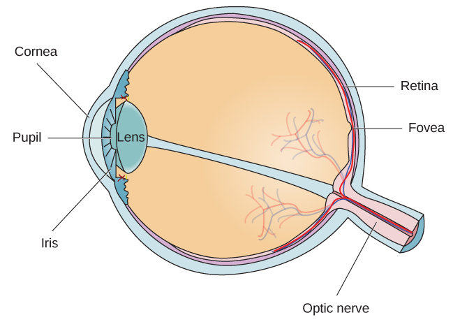
- Vision starts as light waves travel across the cornea and enter the eye through the pupil.
- The cornea is a transparent covering of the eye.
- It is also involved in focusing light waves that enter the eye.
- The pupil is a small opening in the eye
- It can change size depending on the ==amount of light or emotional arousal==.
- Through expansion and constriction of the pupil, it can allow or reduce the amount of light that enters the eye.
- The iris is the colored portion of the eye.
- It has muscles connected to it that control the size of the pupil.
- The cornea is a transparent covering of the eye.
- After light enters the pupil, it crosses the lens.
- The lens is a curved, transparent structure that aid in providing additional focus.
- It is attached to muscles that change the lens' shape to focus light from near or far objects.
- The lens will then focus the images into the fovea.
- The fovea is a small indentation in the retina.
- The retina is a light-sensitive lining at the back of the eye.
- Photoreceptor cells densely packed in the fovea detect light and are converted into nerve signals.
Photoreceptors
-
Different photoreceptors, which can detect light, can be found in the eye.
-
A cone is a photoreceptor that works best in bright light conditions.
- They are very sensitive to detail and provide tremendous spatial resolution.
- They also provide the ability for us to perceive color
- They are concentrated in the fovea, where images tend to be focused.
-
A rod is a photoreceptor that works best in low light conditions
- They lack the spatial resolution or the color function of the cones.
- They are mostly involved in our vision in dimly lit environments.
- They are also involved in our perception of movement in the periphery of our visual field.
-
There is a delay in transitioning between cone and rod activity.
- Night blindness is a condition where your rods do not transform light as efficiently as they could.
How Visual Information is Processed
- Photoreceptors, such as rods and cones, are connected via the retinal ganglion cells.
- Axons from retinal ganglion cells converge outwards and exit through the optic nerve.
- Optic nerves from both eyes converge in a point below the brain called optic chiasm.
- Here, visual information from the right eye is sent in the left hemisphere of the brain, and visual information from the left eye is sent in the right hemisphere of the brain.
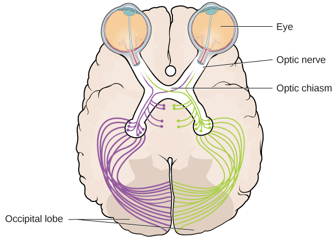
- Once inside the brain, visual information is independently processed in different pathways in the occipital lobe called the what pathway and the why/how pathway.
- The what pathway is involved in object recognition and identification.
- The where/how pathway is involved with location or movement in space and how might one interact with the visual stimulus.
Blind Spots
- These are points in the visual field that we are not consciously aware of.
- There are some reasons why we are not consciously aware of it:
- Blind spots from either eye do not overlap.
- This is because each eye has a slightly different view of the visual field.
- Our visual system fills in the visual information in the blind spot.
- This is why although we cannot respond to visual information that occurs in that field, we are not also aware that information is missing.
- Blind spots from either eye do not overlap.
Color Perception
- There are theories in place that try to explain how we perceive color
Trichromatic Theory of Color Vision
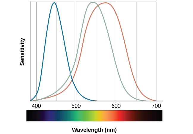
- The human eye has three cones that mediate color vision.
- Each cone type is very sensitive to a specific wavelength of light.
- The trichromatic theory of color vision tells us that each cone type is receptive to red, green, and blue.
- Each color in the spectrum can be produced by combining different amounts of red, green, and blue.
Opponent-Process Theory
- Opponent-Process Theory says that color is coded in opponent pairs.
- These opponent pairs are: black-white, yellow-blue, green-red.
- This theory says that some cells in the visual system are excited by one of the opponent colors and inhibited by the other.
- This means that a cell that is activated by wavelengths associated with yellow, would be inhibited by wavelengths associated with blue, and vice versa.
- This also implies that we do not experience yellowish-blue or greenish-red.
Afterimages
-
It is a phenomena where we continue to receive a visual sensation even after the visual stimulus is removed.
-
Example: When you stare briefly in the sun then look away from it, you may still perceive a spot of light even though the stimulus (in this case, the sun) has been removed.
-
Often when color is involved, the other color in an opponent color pair becomes the color of the negative afterimage.
When the image is green, the afterimage becomes red. -
Both theories for color vision are shown to apply to different parts of the nervous system.
- When visual information is processed in the retina, the trichromatic theory applies.
- Once the signal moves into the brain, the cells respond consistent with opponent-process theory
Depth Perception
- The ability to perceive spatial relationships in 3D space is known as depth perception
- We use a variety of cues to establish our sense of depth.
- Cues that rely on two eyes are called binocular cues.
- One example of a binocular cue is binocular disparity.
- This refers to the slightly different view that each of our eyes receive.
- Sometimes, we can also pick up depth even if a stimulus is in 2D (such as in a photograph or painting)
- This is because we rely on monocular cues in this case.
- These are cues that require only one eye.
- Linear perspective is an example of a monocular cue.
- This says that we perceive depth when we see parallel lines that seem to converge in an image.
- This is because we rely on monocular cues in this case.
Stereoblindness
- a condition when a person is unable to respond to binocular depth cues and relied heavily on monocular depth cues.